Neuroplasticity is like magic …
Neuroplasticity is the brain's capacity to change shape physically when exposed to something new. It’s a fabulous phenomenon.
That’s why you no longer have to think about how to drive. You can think about what’s on the menu instead of how you’re physically going to drive your car from your home to the restaurant.
It’s how we learn new things, how we gather skills and knowledge—and hopefully wisdom—over our lifetime.
If we do those new things regularly, they become habits - like driving - which is simply the result of neurons working together as a team, sending electrochemical signals to each other often.
You’ve heard that ‘neurons that fire together wire together?’
This is what they do to become a working team - a neural pathway you can rely on to automatically execute anything you’ve done consistently.
Without neuroplasticity, we would never have evolved to where we are today. We’d need checklists to tell us what to do and how to do it because we wouldn’t have the capacity to learn anything new, hold it in our working memory, and transfer it to our long-term memory. And habits couldn’t be formed either.
You'd never have learned anything without the brain's amazing capacity to change shape. People who are severely cognitively challenged are unable to learn, but they’re the exception to this rule.
Each brain is a fingerprint—we become who we are through the way our brains change in response to our unique experiences, which we undergo from before birth and then throughout our lives.
Each of us experiences the world differently and will, therefore, have a brain that reflects these differences in its unique neural pathways.
We didn’t always know about this fabulous capacity, but some researchers had a suspicion that something was happening inside the brain before we could peek inside and see it happening. Here’s a not exhaustive list of a few of them:
• William James was an early, suspicious researcher. In his 1890 book, ‘The Principles of Psychology’, he stated, ' Organic matter, especially nervous tissue, seems endowed with a very extraordinary degree of plasticity.’
• Experiments conducted by Karl Lashley in the 1920s using Rhesus monkeys revealed experience-driven, changed neural pathways in these primates.
• Marian Diamond, a pioneer in neuroplasticity in the 1960s, started her work in this area by examining how rats' brains changed. She was one of the first researchers to publish research on neuroplasticity.
• Also in the 1960s, researchers had examined cases of people who had massive strokes but were able to regain full or partial physical and mental functioning.
• Michael Merzenich has been a pioneer in neuroplasticity for over three decades and was one of the researchers to delineate between childhood and adulthood neuroplasticity.
We now know without a shadow of a doubt that our brains do change shape, which can serve us, but some changes DO NOT.
Before we dive into the Achilles heel of brain change, it’s important to grasp that there are two types of neuroplasticity.
[You can skip the two sections below and move directly to where the magic turns dark, but these two sections lay the foundation for the two different types of neuroplasticity.]
Structural neuroplasticity
This is the type we’ve discussed up until this point - the ability of the brain to change shape due to what it experiences. Although we mostly think about what we do, our actions, or what is done to us in relation to brain change.
However, our brain can also change shape from thoughts we think often, including memories we re-live, which is linked to post-traumatic stress disorder (PTSD).
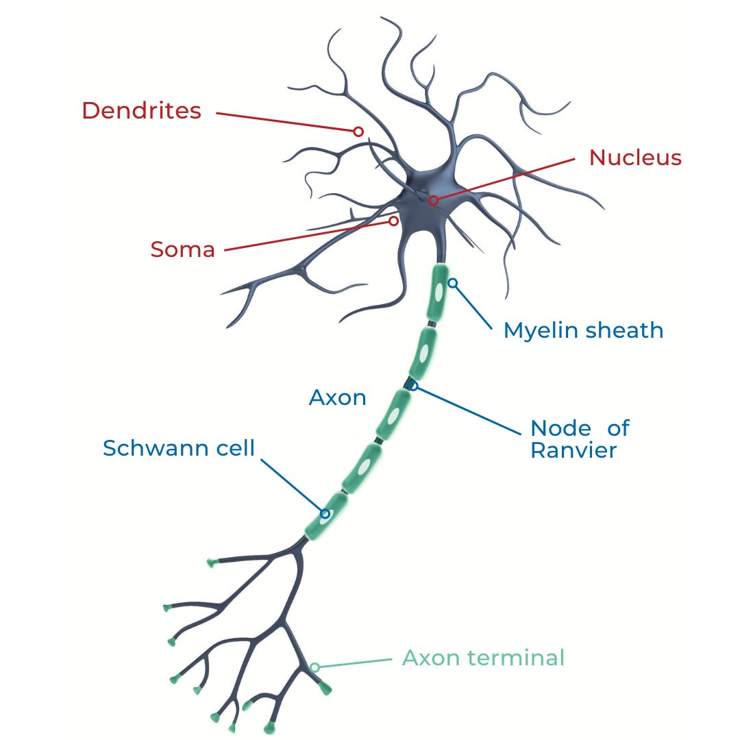
Our brains change shape because the smallest structures, namely axons, dendrites, and synapses, that comprise our specialized 80+ billion neurons, all change when the brain is exposed to new experiences.
All these neuronal structures must change shape to accommodate the information the brain is exposed to.
Functional neuroplasticity
If you’ve ever seen a video or met a person who has had a stroke or suffered a brain injury, this is the type of neuroplasticity they hope comes into play to help them regain lost functionality.
They’ve lost a cognitive and/or physical capacity due to an accident or stroke and then (hopefully) slowly regain the lost capacity.
Researchers used to believe that the functions move from the damaged area to an undamaged area so that functions are restored.
However, some researchers suggest this is not the case and that undamaged or underutilised neurons in the area where the damage occurred are used to restore functionality (Makin TR et al.).
Regardless of type, it feels like magic when we watch a child take its first steps, learn a new word, or when we learn a new skill even as adults. Similarly, it must feel magical when a person thinks they’d lost functionality in an area, and it re-appears.
The following section relates to the first type of neuroplasticity, the structural kind, and how it doesn’t always serve us, especially when we’re young.
Here’s where its magical-like ability turns dark
Very few people know that the Achilles heel of our brain's wonderful capacity to change shape has a dark side.
Firstly, it is far easier to create unhealthy and dangerous habits versus the ones that support our health.
Why? The answer lies deep within the brain's desire to survive and is linked to a neurotransmitter called dopamine (Lembke, A.).
Neurotransmitters are tiny compounds that act like messengers between neurons—they tell them what to do. Some of these messengers are excitatory, i.e., they tell the neurons to do something. Others are inhibitory, i.e., they stop neurons from doing something.
Both types of messengers are important because they both have a role to play in our survival and wellbeing. We have many different types of these messengers, some of which you’d be familiar with, such as serotonin, acetylcholine, and melatonin.
Whenever we do anything that supports our survival dopamine is released. We therefore associate those actions with feeling good and so naturally we repeat them. This is a natural response and not one we can consciously direct.
Think about having an orgasm, finding and eating a satisfying meal when you’re hungry, or being involved in a deep and meaningful conversation with someone that you care about or love. These are dopamine-fueled experiences because they support survival.
It’s why we crave chocolate but never broccoli
Every time we anticipate or do something that supports our survival, the brain releases dopamine.
Dopamine has been called the pleasure neurotransmitter, but it’s also involved in anticipation and motivation because it’s released when we are motivated to do something that we know feels good and when we anticipate the pleasure we’ll experience.
Sex, food, and connection are all activities that lead to a release of dopamine because they all underpin our physical survival, which increase our chances of perpetuating our species.
It’s therefore easier to get into the habit of doing something that leads to pleasure, like eating chocolate, versus broccoli, as less of a brain ‘high’ is produced from healthier habits.
You can see where this is going:
These activities can also all become addictions because the brain gets used to high levels of dopamine and then wants them to stay at that level.
When the level dips, we feel demotivated and miserable. The brain’s natural desire to get back to feeling motivated and happy will drive us to engage in behaviors that keeps dopamine levels high.
This is also why seemingly innocuous activities like drinking coffee, eating chocolate, overeating processed foods, and binge-watching Netflix also release dopamine. They all feel good, and even though they’re not directly linked to survival, they produce a sense of anticipation and pleasure.
Whereas other activities, for example, like eating a bowl of lightly steamed broccoli or meditating, do not release the same amount of dopamine. They don’t support survival - at least not in the short term.
So, we naturally gravitate to activities that release the most amount of dopamine and avoid the ones that lead to a dip in levels.
Technology, especially social media, supports the release of large amounts of dopamine because of the ease of access to stimulating images, ideas, ‘likes’, etc., and how quickly the brain responds to them.
These are also the factors linked to the addictive potential of any behavior or substance.
But before we get to the role that technology and social media play in the developing brain, we need to examine why the developing brain is especially vulnerable to experience.
Periods of significant and rapid neural development, in which the brain is sensitive to less-than optimal experiences and interactions. Image from ‘The impact of developmental timing for stress and recovery’ (2015) by Dylan G. Gee and B.J. Casey; Copyright License: CC BY-NC-ND 4.0
[NOTE: This is an important image so it will appear in Part 2 again.]
A young brain is an opportunistic one …
The ‘windows of neurodevelopmental opportunity’ are so called because the brain is primed to be very malleable—' plastic’—when we are young, and specific periods of time act like ‘windows’ for specific learning activities to occur.
In our youth, our brains are especially focused on adapting and adjusting neuronal connections and forming new ones in relation to the environment.
Why?
Because this allows the brain to quickly adapt to a wide range of environments and experiences that may all be important for its survival.
The early years of a child’s life are ones of rapid growth for the body and brain. Researchers have found that at birth, each child’s neuron in the cerebral cortex has about 2,500 synapses. These are the tiny gaps between neurons over which neurotransmitters must jump to move messages between them.
By age three, each neuron now has about 15,000 synapses, but the average adult neuron only has about 7,500.
So, what’s happened?
The developing brain undergoes a process called ‘synaptic pruning’ in which synapses that aren’t used as regularly (or at all) are removed. Apoptosis also occurs, which is a process whereby unused neurons are discarded.
Think about whether you picked up a musical instrument when you were young. If you did and practiced regularly over the years of childhood, your brain kept the connections it was building as it assumed this was an activity you’d continue to perform.
If not, your ability to play a musical instrument became less easy to acquire with each passing year because your brain was focused on strengthening connections between neurons that you were using, such as for playing football or dancing.
In this simple and efficient way, the brain removes connections that aren’t being used and strengthens those that are.
But what happens when the environment isn’t optimal for brain development? When connections for optimal development aren’t formed while ones for dysfunction are formed?
Let’s step back in time to gain a broader perspective.
The vulnerable, developing brain - two dictators taught us a lot
In 1989, the Romanian dictator Nicolae Ceausescu was overthrown, and the world discovered that 170,000 children were being raised in Romania's impoverished orphanages.
Researchers Fox, Nelson and Zeanah have been involved in a longitudinal research project wherein they’ve followed a group of children who were originally institutionalised under that regime. (Nelson et al.)
They found that the institutionalized children had neurological delays in cognitive function, including language, along with lags in motor development.
They also exhibited deficits in socio-emotional behaviors, had more psychiatric disorders, and showed changes in the patterns of electrical activity in their brains, as measured by EEG, compared to the control group of children.
However, children moved into foster care showed improvements in intellectual capacity, language, and social-emotional functioning. They were also able to form secure emotional-attachment relationships with their caregivers, and their ability to express emotions improved.
Unfortunately, despite these improvements, they still lagged behind compared to the non-institutionalised children in the control group. However, those who’d moved to foster care before age two improved the most and by age eight, their EEGs were no different from those in the control group.
MRIs (to reveal structural differences) used on the institutionalised children revealed smaller brains, less grey matter (neurons), and less white matter (myelin) compared to the control group. The institutionalized children who moved into foster homes recovered some of the missing white matter volume over time, but their gray matter volume stayed low, regardless of whether they’d been moved into stable foster homes (Sheridan, MA).
In other words, some of the neurophysiological changes were permanent, and changing their circumstances didn’t reverse the damage. (Sheridan MA et al; Laine-Frigren T.)
Another study, which started after the Second World War (WW2), revealed that children separated from both caregivers during evacuations all had higher average salivary cortisol and plasma adrenocorticotropic hormone (ACTH) concentrations than the control group.
Interestingly, women had higher baseline levels of both compounds compared to their male counterparts, even 60 years later. (Pesonen A-K, et al.)
Researchers are of the opinion that the amygdala, which is a paired structure within our temporal lobe, amd involved with emotion processing, develops differently when children are exposed to early life stress (ELS).
This impacts how they respond to stress across their lifetime. (Fareri DS et al.)
[Note: I use the term technology and tech-immersion along with social media because iPads that are given to pre-verbal children and online pornography aren’t social media, but still impact the developing brain, although a lot of the current research is focused on the neurological effects of social media.]
In Part 2 we take a deeper dive into exactly why online technology and smartphones are so mesmerising to the developing brain, especially as it enters puberty, and why this matters for the future. The reference list is also in Part 2.


